The Anatomy of the Ring: Understanding Ligament and Joint Trauma
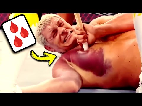
In the high-impact world of the WWE, the line between choreographed entertainment and physical catastrophe is often razor-thin. When examining the knee injury of Logan Paul, we see a classic example of mechanical failure under extreme stress. The medial collateral ligament (MCL) is the primary stabilizer on the inner side of the knee, connecting the femur to the tibia. During a high-altitude maneuver like a 'frog splash,' any slight misalignment upon landing can cause the joint to buckle inward, leading to what clinicians call valgus stress. A grade three MCL tear represents a complete rupture, which typically results in significant joint laxity and immediate loss of athletic function.
Assessing these injuries in an acute setting is notoriously difficult. The primary challenge is that pain and swelling often mask the true extent of the damage. In a clinical exam, a physician looks for laxity—the lack of a firm endpoint when stressing the ligament—compared to the healthy side. If the joint opens up without resistance, the ligament is no longer providing support. Frequently, these injuries do not occur in isolation. They often present as the 'terrible triad,' involving the MCL, the ACL (anterior cruciate ligament), and the meniscus. This combination significantly complicates the recovery timeline and often shifts the prognosis from simple rehabilitation to surgical necessity.
| Injury Grade | Clinical Presentation | Recovery Pathway |
|---|---|---|
| Grade 1 | Micro-tears, minimal swelling | 1-2 weeks of rest |
| Grade 2 | Partial tear, moderate laxity | 3-6 weeks with bracing |
| Grade 3 | Complete rupture, total instability | 6+ months or surgery |
When we transition to upper extremity trauma, such as the shoulder separation experienced by Randy Orton, we must distinguish between a dislocation and a separation. A shoulder dislocation involves the humerus popping out of the glenoid socket, whereas a shoulder separation involves the AC joint (acromioclavicular joint). This joint connects the shoulder blade to the clavicle. A high-impact fall directly onto the shoulder can tear the ligaments holding these bones together, creating a visible bump. This type of injury is graded from one to six, with higher grades indicating that the clavicle has been displaced significantly from its original position.
Facial Impact and Neurological Risks: From Broken Noses to Brain Health

Facial injuries in wrestling are often dismissed as superficial, but the medical reality is far more complex. When John Cena suffered a severely deformed nose during a match, the primary concern was not just the aesthetic fracture of the nasal bones. A physician's first priority in such cases is to rule out a septal hematoma. This occurs when blood pools between the nasal septum and its overlying perichondrium. If left untreated, the pressure from the blood can cut off the blood supply to the cartilage, leading to necrosis and a permanent 'saddle nose' deformity. Furthermore, any severe facial trauma carries the risk of a CSF (cerebrospinal fluid) leak, indicating a breach in the barrier between the brain and the external environment.
Impacts to the cheek and eye area, as seen with Bianca Belair and Sasha Banks, carry the risk of fracturing the zygomatic arch. While simple bruising, known as ecchymosis, is common, certain patterns of discoloration serve as red flags. For instance, 'raccoon eyes' (bilateral periorbital ecchymosis) or 'battle sign' (bruising behind the ear) are classic indicators of a basilar skull fracture. These are medical emergencies that suggest the base of the skull has been compromised, potentially affecting cranial nerves or leading to intracranial bleeding.
ここからが大事な
ポイントです
具体例・注意点・明日から使えるヒントを整理しています。
✨無料閲覧で全文 + 図解の完全版を3日間いつでも読み返せる
あなたの好きな動画も、
1分でAI要約
📚 お気に入り保存 + ✨ あなたの動画をAI要約
(無料登録10秒)
✏️ この記事で学べること
- ▸、 。 、 、 。 、 。
10秒で完了・パスワード作成不要