The Biological Mechanics of Iris Pigmentation and Light

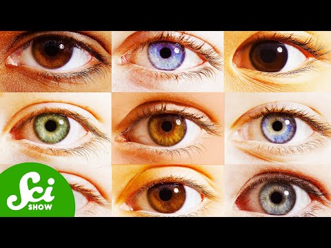
The human eye is often considered a static feature, yet its color is the result of complex biological structures interacting with light. The iris serves as the gateway for light entering the eye, utilizing muscles to adjust the pupil's size. Within the iris, two distinct layers hold the secret to your specific shade. Most of the focus lies on melanocytes, cells found in both layers that produce melanin. This is the same pigment responsible for skin and hair color. Melanin's primary function in the eye is to absorb light; the more melanin present, the darker the eye appears, as less light is reflected back to the observer.
Interestingly, the perception of color in lighter eyes, such as blue or green, is not due to blue or green pigments. Instead, it is a result of low melanin levels allowing short-wavelength light to reflect off collagen fibers in the iris stroma. This phenomenon is similar to why the sky appears blue. There are two main forms of melanin: a black or dark brown variety, and a reddish-yellow variety. The specific ratio and density of these pigments determine the final hue, much like mixing paints on a palette.
| Pigment Type | Common Eye Colors | Light Interaction |
|---|---|---|
| High Eumelanin | Brown, Black | High absorption, low reflection |
| Low Eumelanin | Blue, Gray | High scattering of short wavelengths |
| Pheomelanin | Green, Amber | Selective reflection of yellow/red tones |
Developmental Shifts and the Impact of Aging
The journey of eye color begins in the womb, but it rarely ends there. Many infants are born with lighter eyes because their melanocytes have not yet moved into their final positions or begun producing full levels of melanin. Over the first few years of life, as these cells mature, the iris typically darkens. Studies on twins have shown that for about 10% to 15% of the Caucasian population, this darkening process can continue well into adulthood, suggesting a strong genetic link to age-related color transitions.
Conversely, as humans reach senior years, the eyes may appear to lighten or develop new features. A common occurrence is the formation of the Arcus Senilis, a white, gray, or blue-ish ring around the edge of the cornea caused by lipid deposits. While usually benign, these changes can alter the perceived color of the eye, giving it a halo-like effect. It is a reminder that the eye is a living, evolving system rather than a fixed camera lens.
ここからが大事な
ポイントです
具体例・注意点・明日から使えるヒントを整理しています。
✨無料閲覧で全文 + 図解の完全版を3日間いつでも読み返せる
あなたの好きな動画も、
1分でAI要約
📚 お気に入り保存 + ✨ あなたの動画をAI要約
(無料登録10秒)
✏️ この記事で学べること
- ▸、 。 、 、 、 。
10秒で完了・パスワード作成不要